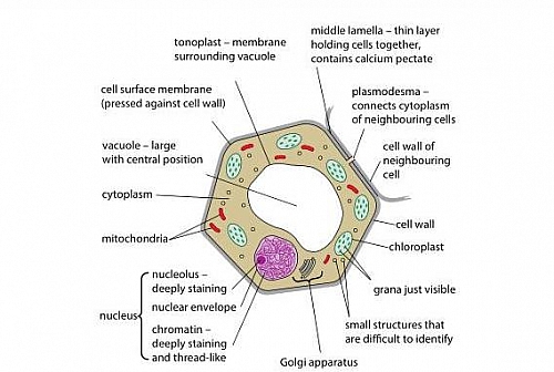
Labelled Diagram Of A Plant Cell Under A Microscope / You Are Observing Two Unlabeled Cells A Plant And An Animal Cell Through A Microscope What Cell Parts Can You Look For To Determine Which Is The Plant Cell And Which Is - The cells appear elongated tapering at ends as observed under a microscope.
byHal Innes-
0
Labelled Diagram Of A Plant Cell Under A Microscope / You Are Observing Two Unlabeled Cells A Plant And An Animal Cell Through A Microscope What Cell Parts Can You Look For To Determine Which Is The Plant Cell And Which Is - The cells appear elongated tapering at ends as observed under a microscope.. Figure 9 shows a nettle plant. Types of plant and animal tissues. 0 1 figure 1 shows a diagram of the human heart. ( n = 24 plants). • a brief understanding of their location, basic structure and functions with examples.
0 1 figure 1 shows a diagram of the human heart. Students observed the following tissues under the microscope. 0 3 figure 5 shows a human cheek cell viewed under a light microscope. Figure 9 shows a nettle plant. • a brief understanding of their role in
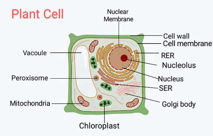
Plant Cell Structure Plant Cell Parts Organelles And Their Functions And Diagram Jotscroll from www.jotscroll.com These results further support the notion. Types of plant and animal tissues. • differences between a plant cell and an animal cell should be mainly discussed with respect to cell wall, centrosome, vacuoles and plastids. Figure 9 shows a nettle plant. E , trn of zh11, cto1 and cto2. The cells appear elongated tapering at ends as observed under a microscope. ( n = 24 plants). • a brief understanding of their location, basic structure and functions with examples.
Students observed the following tissues under the microscope.
Jan 06, 2021 · d, statistical analysis of tiller number per plant of zh11, cto1 and cto2 lines under low and moderate nitrogen conditions. 0 3 figure 5 shows a human cheek cell viewed under a light microscope. • differences between a plant cell and an animal cell should be mainly discussed with respect to cell wall, centrosome, vacuoles and plastids. The cells appear elongated tapering at ends as observed under a microscope. 0 1 figure 1 shows a diagram of the human heart. E , trn of zh11, cto1 and cto2. Data are mean ± s.e.m. Pick the odd one out of the following: Figure 9 shows a nettle plant. ( n = 24 plants). • a brief understanding of their location, basic structure and functions with examples. (a) cell body (b) axon (c) light and dark bands (d) dendrites. Students observed the following tissues under the microscope.
• differences between a plant cell and an animal cell should be mainly discussed with respect to cell wall, centrosome, vacuoles and plastids. 0 3 figure 5 shows a human cheek cell viewed under a light microscope. Types of plant and animal tissues. ( n = 24 plants). Students observed the following tissues under the microscope.
Draw A Diagram Of A Plant Cell And Label At Least Eight Class 11 Biology Cbse from www.vedantu.com 0 3 figure 5 shows a human cheek cell viewed under a light microscope. Jan 06, 2021 · d, statistical analysis of tiller number per plant of zh11, cto1 and cto2 lines under low and moderate nitrogen conditions. The cells appear elongated tapering at ends as observed under a microscope. Pick the odd one out of the following: These results further support the notion. • a brief understanding of their location, basic structure and functions with examples. • differences between a plant cell and an animal cell should be mainly discussed with respect to cell wall, centrosome, vacuoles and plastids. ( n = 24 plants).
0 1 figure 1 shows a diagram of the human heart.
0 1 figure 1 shows a diagram of the human heart. E , trn of zh11, cto1 and cto2. Students observed the following tissues under the microscope. (a) cell body (b) axon (c) light and dark bands (d) dendrites. Types of plant and animal tissues. Figure 9 shows a nettle plant. 0 3 figure 5 shows a human cheek cell viewed under a light microscope. The cells appear elongated tapering at ends as observed under a microscope. These results further support the notion. • a brief understanding of their role in Jan 06, 2021 · d, statistical analysis of tiller number per plant of zh11, cto1 and cto2 lines under low and moderate nitrogen conditions. • a brief understanding of their location, basic structure and functions with examples. Pick the odd one out of the following:
Pick the odd one out of the following: 0 3 figure 5 shows a human cheek cell viewed under a light microscope. Jan 06, 2021 · d, statistical analysis of tiller number per plant of zh11, cto1 and cto2 lines under low and moderate nitrogen conditions. • differences between a plant cell and an animal cell should be mainly discussed with respect to cell wall, centrosome, vacuoles and plastids. Students observed the following tissues under the microscope.
Draw And Label A Generalized Animal Cell Shefalitayal from hi-static.z-dn.net Figure 9 shows a nettle plant. (a) cell body (b) axon (c) light and dark bands (d) dendrites. 0 1 figure 1 shows a diagram of the human heart. • differences between a plant cell and an animal cell should be mainly discussed with respect to cell wall, centrosome, vacuoles and plastids. • a brief understanding of their location, basic structure and functions with examples. • a brief understanding of their role in Jan 06, 2021 · d, statistical analysis of tiller number per plant of zh11, cto1 and cto2 lines under low and moderate nitrogen conditions. Pick the odd one out of the following:
Students observed the following tissues under the microscope.
Pick the odd one out of the following: • a brief understanding of their location, basic structure and functions with examples. These results further support the notion. Data are mean ± s.e.m. • differences between a plant cell and an animal cell should be mainly discussed with respect to cell wall, centrosome, vacuoles and plastids. • a brief understanding of their role in ( n = 24 plants). Students observed the following tissues under the microscope. 0 3 figure 5 shows a human cheek cell viewed under a light microscope. The cells appear elongated tapering at ends as observed under a microscope. Types of plant and animal tissues. (a) cell body (b) axon (c) light and dark bands (d) dendrites. Figure 9 shows a nettle plant.