Plant Cell Under Microscope Labeled - Mitosis Through The Microscope Advances In Seeing Inside Live Dividing Cells Science - Jul 25, 2021 · general features of plant cell.
byHal Innes-
0
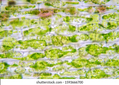
Plant Cell Under Microscope Labeled - Mitosis Through The Microscope Advances In Seeing Inside Live Dividing Cells Science - Jul 25, 2021 · general features of plant cell.. Here the axonal bundles of the dorsal root ganglions are imaged by fluorescence line scanning confocoal microscope (zeiss lsm710) equipped with multiple lasers. Just under the rigid cell wall is the more fluid cell membrane. (d) electron microscopic image of a pancreatic beta cell. Explore topics on usage, care, terminology and then interact with a fully functional, virtual micro Under the microscope, it shows many different parts.
(d) electron microscopic image of a pancreatic beta cell. Jul 01, 2021 · figure created with biorender.com. Microscope slides preparation styles and techniques using prepared microscope slides. Under the microscope, it shows many different parts. Jul 25, 2021 · general features of plant cell.
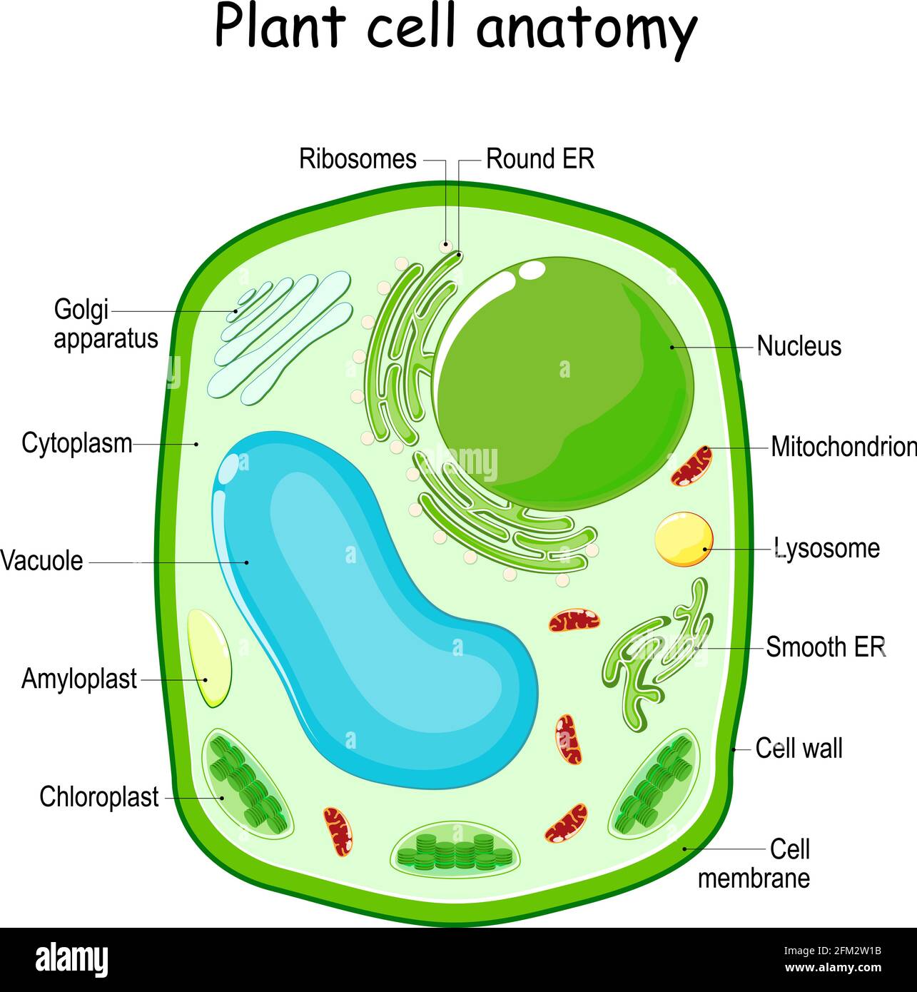
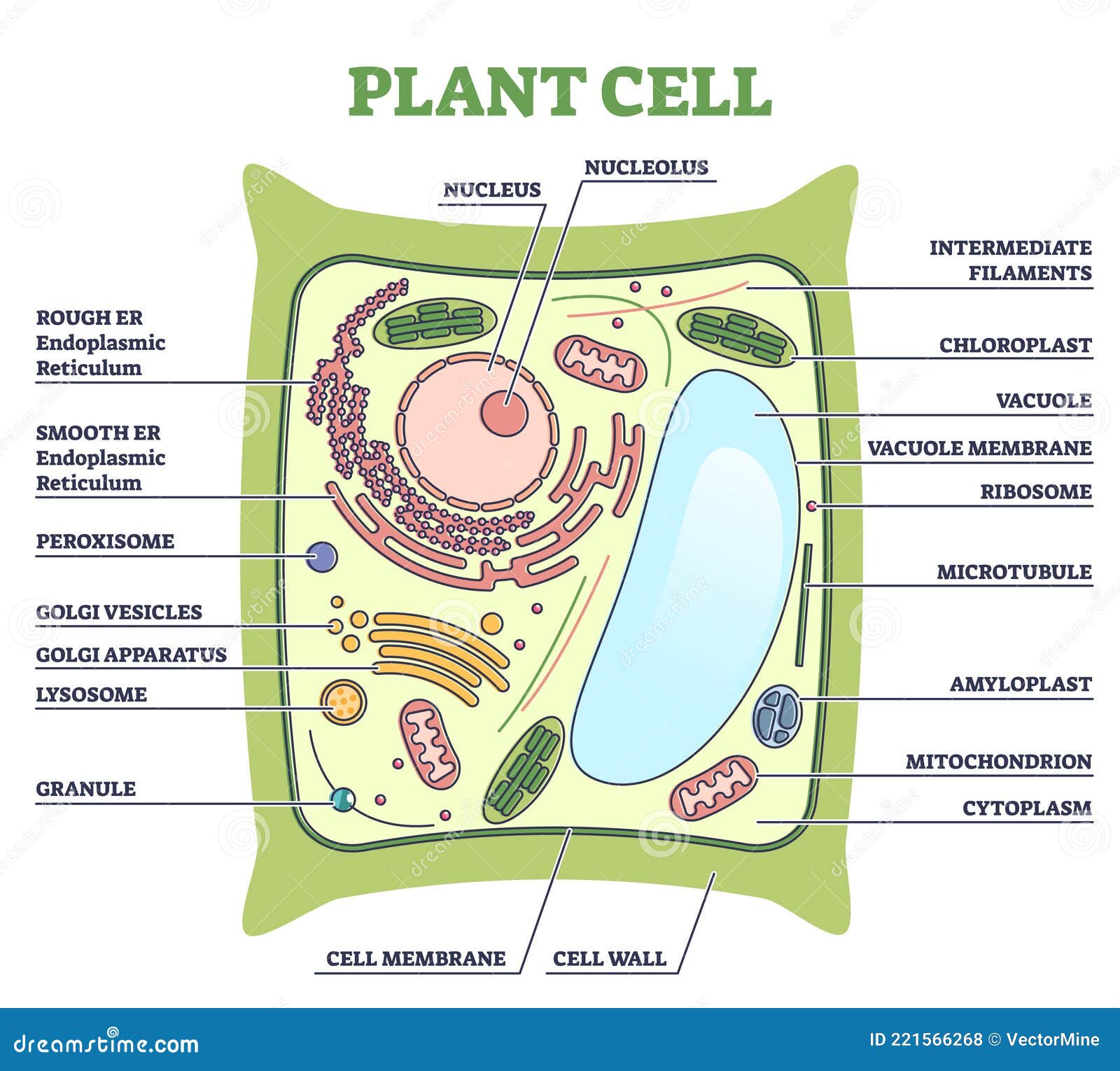
Plant Cell Nucleus High Resolution Stock Photography And Images Alamy from c8.alamy.com The cytoplasm enclosed within the cell membrane does not exhibit much structure when viewed by electron microscopy. Use the following animation to explore bacterial structure. Plants are made up two structural systems i.e the shoot system and the root system, whereby the shoot system is made up of structures that ie above the ground including leaves, stems, fruits, flowers while the root system is made up of roots, tubers and rhizobial structure that lie below the ground and its the origin of growth of plants. Microscope slides preparation styles and techniques using prepared microscope slides. Jul 04, 2020 · a model of a typical plant cell is found to be rectangular in shape, ranging in size from 10 to 100 µm. Under the microscope, it shows many different parts. There are three structural parts of the microscope i.e. Explore topics on usage, care, terminology and then interact with a fully functional, virtual micro
Microscope slides preparation styles and techniques using prepared microscope slides.
Here the axonal bundles of the dorsal root ganglions are imaged by fluorescence line scanning confocoal microscope (zeiss lsm710) equipped with multiple lasers. The beta cell senses the increased. Diagram of parts of a microscope. (b) looking at a pancreatic islet with h&e (haemotoxylin and eosin) staining under a compound microscope. (d) electron microscopic image of a pancreatic beta cell. They have an outer cell wall that gives them shape. Under the microscope, it shows many different parts. Jul 25, 2021 · general features of plant cell. Jul 04, 2020 · a model of a typical plant cell is found to be rectangular in shape, ranging in size from 10 to 100 µm. Microscope slides preparation styles and techniques using prepared microscope slides. Just under the rigid cell wall is the more fluid cell membrane. There are three structural parts of the microscope i.e. Plants are made up two structural systems i.e the shoot system and the root system, whereby the shoot system is made up of structures that ie above the ground including leaves, stems, fruits, flowers while the root system is made up of roots, tubers and rhizobial structure that lie below the ground and its the origin of growth of plants.
Here the axonal bundles of the dorsal root ganglions are imaged by fluorescence line scanning confocoal microscope (zeiss lsm710) equipped with multiple lasers. Under the microscope, it shows many different parts. (b) looking at a pancreatic islet with h&e (haemotoxylin and eosin) staining under a compound microscope. The cytoplasm enclosed within the cell membrane does not exhibit much structure when viewed by electron microscopy. Jul 25, 2021 · general features of plant cell.
Plant Cell Labeled Stock Illustrations 75 Plant Cell Labeled Stock Illustrations Vectors Clipart Dreamstime from thumbs.dreamstime.com Use the following animation to explore bacterial structure. Jul 04, 2020 · a model of a typical plant cell is found to be rectangular in shape, ranging in size from 10 to 100 µm. The cytoplasm enclosed within the cell membrane does not exhibit much structure when viewed by electron microscopy. The beta cell senses the increased. (d) electron microscopic image of a pancreatic beta cell. (b) looking at a pancreatic islet with h&e (haemotoxylin and eosin) staining under a compound microscope. Plants are made up two structural systems i.e the shoot system and the root system, whereby the shoot system is made up of structures that ie above the ground including leaves, stems, fruits, flowers while the root system is made up of roots, tubers and rhizobial structure that lie below the ground and its the origin of growth of plants. Here the axonal bundles of the dorsal root ganglions are imaged by fluorescence line scanning confocoal microscope (zeiss lsm710) equipped with multiple lasers.
Microscope slides preparation styles and techniques using prepared microscope slides.
Microscope slides preparation styles and techniques using prepared microscope slides. Diagram of parts of a microscope. Each part, known as an organelle, works together to keep the cell functional. Here the axonal bundles of the dorsal root ganglions are imaged by fluorescence line scanning confocoal microscope (zeiss lsm710) equipped with multiple lasers. Under the microscope, it shows many different parts. Just under the rigid cell wall is the more fluid cell membrane. The beta cell senses the increased. Jul 25, 2021 · general features of plant cell. Jul 04, 2020 · a model of a typical plant cell is found to be rectangular in shape, ranging in size from 10 to 100 µm. They have an outer cell wall that gives them shape. Use the following animation to explore bacterial structure. Explore topics on usage, care, terminology and then interact with a fully functional, virtual micro (d) electron microscopic image of a pancreatic beta cell.
Under the microscope, it shows many different parts. (d) electron microscopic image of a pancreatic beta cell. Plants are made up two structural systems i.e the shoot system and the root system, whereby the shoot system is made up of structures that ie above the ground including leaves, stems, fruits, flowers while the root system is made up of roots, tubers and rhizobial structure that lie below the ground and its the origin of growth of plants. Explore topics on usage, care, terminology and then interact with a fully functional, virtual micro Jul 25, 2021 · general features of plant cell.
Plant Cell Under Microscope Images Stock Photos Vectors Shutterstock from image.shutterstock.com Explore topics on usage, care, terminology and then interact with a fully functional, virtual micro Each part, known as an organelle, works together to keep the cell functional. Microscope slides preparation styles and techniques using prepared microscope slides. They have an outer cell wall that gives them shape. Diagram of parts of a microscope. Use the following animation to explore bacterial structure. Jul 04, 2020 · a model of a typical plant cell is found to be rectangular in shape, ranging in size from 10 to 100 µm. Just under the rigid cell wall is the more fluid cell membrane.
Jul 25, 2021 · general features of plant cell.
Jul 01, 2021 · figure created with biorender.com. Diagram of parts of a microscope. Each part, known as an organelle, works together to keep the cell functional. The cytoplasm enclosed within the cell membrane does not exhibit much structure when viewed by electron microscopy. Under the microscope, it shows many different parts. The beta cell senses the increased. Jul 25, 2021 · general features of plant cell. There are three structural parts of the microscope i.e. Plants are made up two structural systems i.e the shoot system and the root system, whereby the shoot system is made up of structures that ie above the ground including leaves, stems, fruits, flowers while the root system is made up of roots, tubers and rhizobial structure that lie below the ground and its the origin of growth of plants. Jul 04, 2020 · a model of a typical plant cell is found to be rectangular in shape, ranging in size from 10 to 100 µm. Microscope slides preparation styles and techniques using prepared microscope slides. Explore topics on usage, care, terminology and then interact with a fully functional, virtual micro (d) electron microscopic image of a pancreatic beta cell.